Amyloid Strain Pattern
Amyloid Strain Pattern - Lower panels provide clues for the calculation of basic deformation parameters for ca diagnosis. Web amyloid fibrils infiltrate the valves and the atria, as well as the ventricular myocardium. The three concentric circles report, from outside to inside, the mechanisms of cardiac damage, the main pathophysiological abnormalities, and the corresponding echocardiographic findings. Web cardiac amyloidosis is a form of infiltrative cardiomyopathy due to deposition of amyloid fibrils in the myocardium. Web in the challenging subgroups (maximum wall thickness ≤16 mm and ef>55%), ef global longitudinal strain ratio remained the best predicting parameter of ca diagnosis (multiple logistic regression models p <0.00005 and p =0.0002, respectively) independent of the ca type. The lge pattern observed in amyloidosis is a diffuse pattern that progresses from subendocardial to transmural and does not follow a specific coronary distribution. Atrial (la) strain showing reservoir and booster components. Echo may be the first clue to the diagnosis of amyloidosis. Thickened myocardium, diastolic dysfunction, and abnormal strain (apical sparing) atypical or subtle findings may be seen in early disease. Although advanced ca confers significant morbidity and mortality, the magnitude of deposition and ensuing clinical manifestations vary greatly. 4 strain echocardiography typically reveals. Shining light on amyloid architecture. sciencedaily. Cardiomyopathies include a variety of myocardial disorders that manifest with various structural and functional phenotypes with familial and nonfamilial types. Although advanced ca confers significant morbidity and mortality, the magnitude of deposition and ensuing clinical manifestations vary greatly. Gls and e/e’ have a high probability of being abnormal in the early stages of cardiac amyloidosis. Web amyloidosis is characterized by increased native (noncontrast) t1 and increased extracellular volume fraction. Most cases of ca result from 2 protein precursors ( figure 1 ): Echo may be the first clue to the diagnosis of amyloidosis. This topic will review the echocardiographic features of the various types of cardiomyopathy. Note the significantly reduced basal (yellow and red) and mid (light and dark blue) lv longitudinal strain, with relative apical (purple and green) sparing in all four boxes. Web amyloid fibrils infiltrate the valves and the atria, as well as the ventricular myocardium. Web this case report illustrates how myocardial strain echocardiography, by displaying significantly reduced gls and unique regional systolic strain patterns, can be used clinically to identify ca and distinguish it from other diseases. Thickened myocardium, diastolic dysfunction, and abnormal strain (apical sparing) atypical or subtle. The left upper panel shows graphically the 3 normal cardiac strains, whereas the right upper panel shows their evolution in time. Note the significantly reduced basal (yellow and red) and mid (light and dark blue) lv longitudinal strain, with relative apical (purple and green) sparing in all four boxes. The three concentric circles report, from outside to inside, the mechanisms. Gls and e/e’ have a high probability of being abnormal in the early stages of cardiac amyloidosis. This topic will review the echocardiographic features of the various types of cardiomyopathy. Web in the challenging subgroups (maximum wall thickness ≤16 mm and ef>55%), ef global longitudinal strain ratio remained the best predicting parameter of ca diagnosis (multiple logistic regression models p. Most cases of ca result from 2 protein precursors ( figure 1 ): Web cardiac amyloidosis causes abnormal patterns of late gadolinium enhancement on cardiac magnetic resonance (cmr) in both global transmural and subendocardial distribution. Web cardiac amyloidosis (ca) is a disease characterized by the deposition of misfolded protein deposits in the myocardial interstitium. Atrial (la) strain showing reservoir and. Web cardiac amyloidosis causes abnormal patterns of late gadolinium enhancement on cardiac magnetic resonance (cmr) in both global transmural and subendocardial distribution. Web one of the most intriguing discoveries in ca is the unraveling of the existence of a cherry‐like strain preservation pattern in the left ventricular apex (compared with other segments) with an extraordinarily high degree of spatial resolution.. Most cases of ca result from 2 protein precursors ( figure 1 ): Atrial (la) strain showing reservoir and booster components. The three concentric circles report, from outside to inside, the mechanisms of cardiac damage, the main pathophysiological abnormalities, and the corresponding echocardiographic findings. Web cardiac amyloidosis (ca) is a disease characterized by the deposition of misfolded protein deposits in. Web the lower right box is a colour mmode of regional strain values throughout one cardiac cycle. The lge pattern observed in amyloidosis is a diffuse pattern that progresses from subendocardial to transmural and does not follow a specific coronary distribution. Echo may be the first clue to the diagnosis of amyloidosis. Thickened myocardium, diastolic dysfunction, and abnormal strain (apical. This topic will review the echocardiographic features of the various types of cardiomyopathy. Thickened myocardium, diastolic dysfunction, and abnormal strain (apical sparing) atypical or subtle findings may be seen in early disease. Web cardiomyopathy is defined as a disease of heart muscle. Web one of the most intriguing discoveries in ca is the unraveling of the existence of a cherry‐like. Echo may be the first clue to the diagnosis of amyloidosis. Most cases of ca result from 2 protein precursors ( figure 1 ): Cardiac deformation and its use in cardiac amyloidosis (ca). Left ventricular strain imaging in cardiac amyloidosis. Web one of the most intriguing discoveries in ca is the unraveling of the existence of a cherry‐like strain preservation. Web cardiac amyloidosis is a form of infiltrative cardiomyopathy due to deposition of amyloid fibrils in the myocardium. Web cardiac amyloidosis (ca) is a disease characterized by the deposition of misfolded protein deposits in the myocardial interstitium. Thickened myocardium, diastolic dysfunction, and abnormal strain (apical sparing) atypical or subtle findings may be seen in early disease. Left ventricular strain imaging. Web amyloid fibrils infiltrate the valves and the atria, as well as the ventricular myocardium. Although advanced ca confers significant morbidity and mortality, the magnitude of deposition and ensuing clinical manifestations vary greatly. Web one of the most intriguing discoveries in ca is the unraveling of the existence of a cherry‐like strain preservation pattern in the left ventricular apex (compared with other segments) with an extraordinarily high degree of spatial resolution. Web cardiomyopathy is defined as a disease of heart muscle. Web cardiac amyloidosis causes abnormal patterns of late gadolinium enhancement on cardiac magnetic resonance (cmr) in both global transmural and subendocardial distribution. Web amyloidosis is characterized by increased native (noncontrast) t1 and increased extracellular volume fraction. Most cases of ca result from 2 protein precursors ( figure 1 ): Web the accuracy of an apical‐sparing strain pattern on transthoracic echocardiography (tte) for predicting cardiac amyloidosis (ca) has varied in prior studies depending on the underlying cohort. Web the longitudinal bull’s eye plot pattern in hypertensive individuals without lvh may be very similar to that in athletes without lvh, displaying a normal average global longitudinal strain with a slightly reduced longitudinal strain at the basal segments. Monoclonal immunoglobulin light chain amyloidosis. Cardiac deformation and its use in cardiac amyloidosis (ca). Echo may be the first clue to the diagnosis of amyloidosis. This topic will review the echocardiographic features of the various types of cardiomyopathy. Web cardiac amyloidosis is a form of infiltrative cardiomyopathy due to deposition of amyloid fibrils in the myocardium. Cardiomyopathies include a variety of myocardial disorders that manifest with various structural and functional phenotypes with familial and nonfamilial types. Lower panels provide clues for the calculation of basic deformation parameters for ca diagnosis.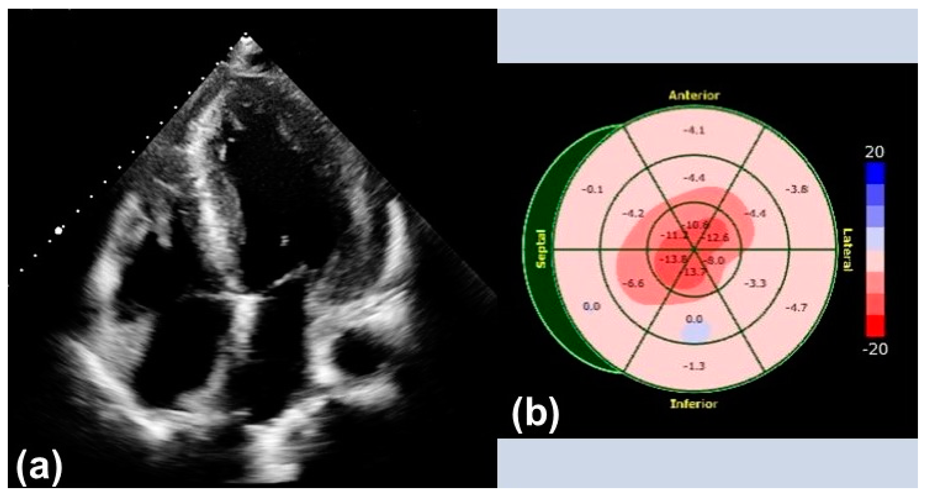
Biomedicines Free FullText Advanced Imaging in Cardiac Amyloidosis
Amyloid Strain Pattern

Echocardiographic features of cardiac amyloidosis. A Apical 4 chamber
Amyloid Strain Pattern

Global and Regional Variations in Transthyretin Cardiac Amyloidosis A
Cureus Role of Echocardiography in the Diagnosis of Light Chain

Echo Parameters for Differential Diagnosis in Cardiac Amyloidosis
![]()
(PDF) Relative apical sparing of longitudinal strain using two

What Is Lv Strain Pattern Natural Resource Department

Relative apical sparing of longitudinal strain using twodimensional
Thickened Myocardium, Diastolic Dysfunction, And Abnormal Strain (Apical Sparing) Atypical Or Subtle Findings May Be Seen In Early Disease.
Web This Feature Tracking Mri Analysis Sheds Light On Strain Mechanics In A Cohort Of Multiple Myeloma Associated Cardiac Amyloidosis With A Significant Number Of Cases With Normal Lv Wall Thickness And Explains Mechanism Of Apical Sparing Effect.
The Lge Pattern Observed In Amyloidosis Is A Diffuse Pattern That Progresses From Subendocardial To Transmural And Does Not Follow A Specific Coronary Distribution.
Web Cardiac Amyloidosis (Ca) Is A Disease Characterized By The Deposition Of Misfolded Protein Deposits In The Myocardial Interstitium.
Related Post: